


The scan comes back with a new 8mm solid nodule in the contralateral lobe. The patient finished lobectomy for stage II NSCLC fourteen months ago. The margins were clean. The adjuvant chemo is done. And now this.
Is it metastatic disease? A second primary? A granuloma? The imaging alone will not tell you. And the decision you make in the next thirty days will determine whether this patient gets curative-intent surgery, systemic therapy, or active surveillance.
This is the most common hard call in thoracic oncology, and it is getting more common every year. As more lung cancer patients reach the two-year and five-year marks, more are walking into follow-up appointments with new nodules. The clinical frameworks for handling them have not kept pace.
The Scale Is Larger Than Most Clinicians Realize
As of January 2025, more than 680,000 Americans were living with a prior lung cancer diagnosis. The National Cancer Institute estimates another 230,000 patients will be diagnosed in 2026. A meaningful fraction have survived long enough to be at risk for exactly this scenario: a new nodule appearing during surveillance imaging.
The numbers on second primary lung cancer are sobering:
Among early-stage survivors, the annual risk of a second primary lung cancer is estimated at 1 to 3.4 percent per year.
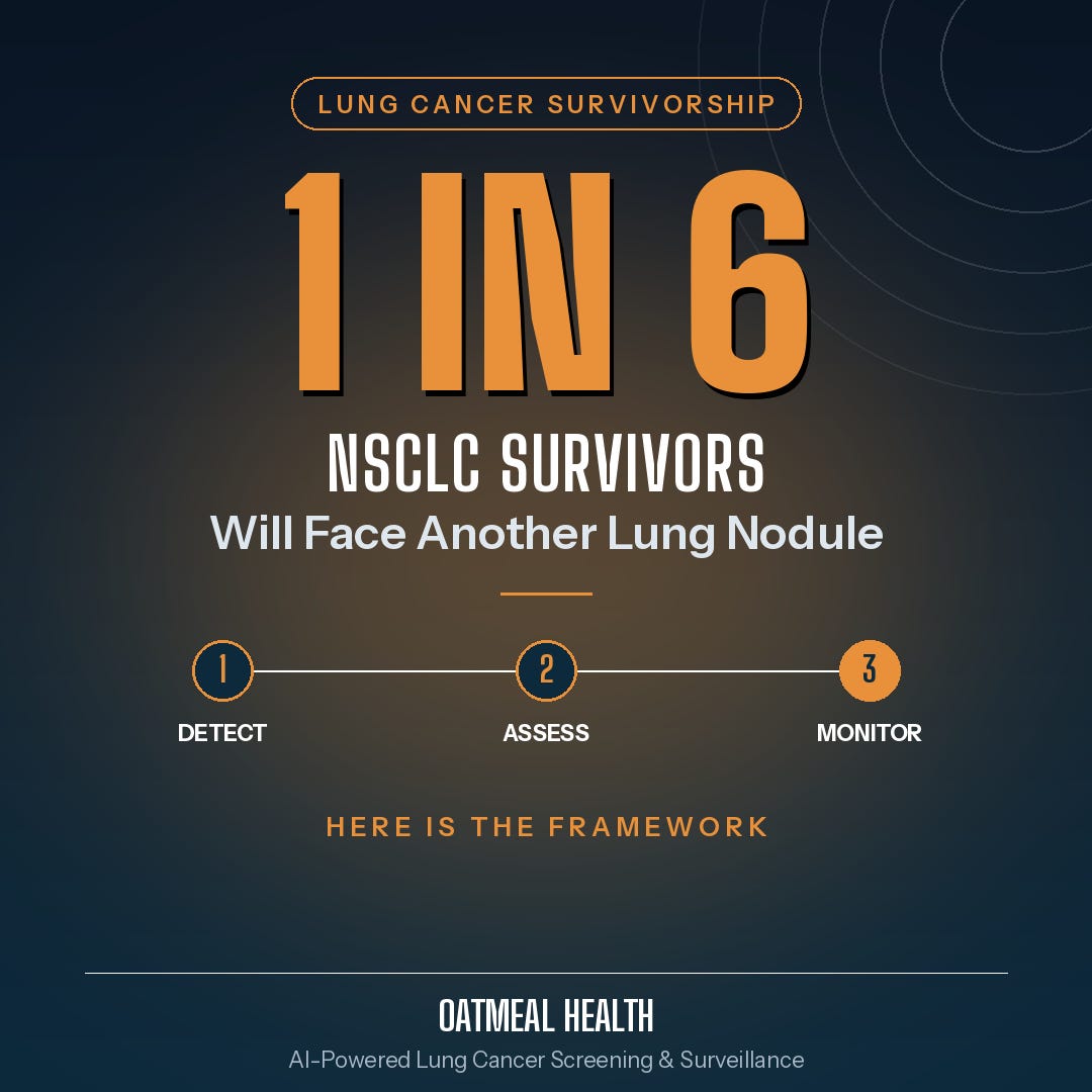
Over five years, the cumulative incidence of a second primary lung cancer in NSCLC survivors approaches 15.9 percent: roughly one in six five-year survivors.
The median time from initial treatment to second primary lung cancer is 25.4 months, right in the middle of the most intensive surveillance window.
For resected stage Ib and II NSCLC patients, 46 percent experienced recurrence within two years of surgery.
If you follow ASCO surveillance guidelines (CT every six months for two years, then annually), you will see nodules. Many will be benign. Some will not be. Every one requires a structured clinical decision.
Why This Call Is Hard
The central challenge is distinguishing three possibilities that imaging often cannot reliably separate: local or distant recurrence of the original NSCLC (which changes treatment goals from curative to palliative in most cases), a second primary lung cancer (which may be an entirely separate, curable tumor if caught early), and benign findings such as granuloma, scar, or infectious nodule.
Standard Lung-RADS criteria were designed for screening populations, not for patients with prior lung cancer. A nodule that would be Lung-RADS 2 in a smoker with no cancer history has a very different clinical meaning in a patient who had an EGFR-mutant adenocarcinoma removed fourteen months ago.
A 2023 proposal in Radiology introduced a Modified Lung-RADS designed specifically for patients with previous malignancies, acknowledging that this population needs different risk thresholds. The approach reflects the growing recognition that post-treatment nodule evaluation is its own subspecialty question.
A Five-Step Framework for Post-Treatment Nodule Evaluation
This framework is not a protocol. It is a structured set of questions ensuring the relevant clinical factors are on the table before a management decision is made.
Step 1: Anchor in Timing and Treatment History
The first question is not 'what does this nodule look like?' It is 'what just happened to this patient, and when?'
Key anchors: time since completion of curative-intent treatment, the type of treatment, and original stage. Radiation therapy changes tissue architecture near treated areas for years. What looks like a new nodule may be radiation fibrosis or post-treatment scar.
Timing matters most in the first twelve months. Within the first year after surgery, most new intrathoracic lesions represent recurrence rather than second primary cancer. Beyond two years, second primary becomes a more significant proportion.
Step 2: Characterize the Nodule Systematically
Before tumor biology enters the conversation, imaging features should be documented in structured form:
Size and growth rate: ASCO surveillance guidelines recommend applying Lung-RADS 1.1 criteria for new nodules at and after the two-year mark. Volume doubling time below 400 days is concerning; above 600 days is reassuring.
Morphology: Solid vs. ground-glass vs. part-solid. Pure ground-glass nodules have a lower probability of being metastatic disease; they are more likely to represent adenocarcinoma in situ or a second primary.
Location: Contralateral nodules, particularly with different morphology from the original tumor, raise the index of suspicion for second primary.
PET avidity: Sensitivity for detecting recurrence or second primary cancer exceeds 98 percent. A new FDG-avid lesion demands tissue-level evaluation unless there is clear clinical contraindication.
Step 3: Review the Biology of the Original Tumor
The molecular and histologic fingerprint of the original cancer informs how to interpret the new lesion.
Histology match: If the original tumor was squamous cell carcinoma and the new nodule shows adenocarcinoma features on imaging, the probability of second primary increases substantially.
Driver mutations: EGFR-mutant adenocarcinoma tends to produce contiguous or lymphatic spread rather than discrete new nodules in early recurrence. Molecular context matters for what workup follows.
Original staging: N2 or N3 disease at original diagnosis substantially raises the prior probability that any new finding represents metastatic disease.
Prior ctDNA testing: If the patient was ctDNA-negative at treatment completion, a new nodule accompanied by a rising ctDNA signal should be treated with high suspicion for recurrence.
Step 4: Apply Risk Stratification to Guide the Workup
With imaging characterization and biological context in hand, the clinical team can apply structured risk assessment to determine workup intensity.
High-risk features that favor biopsy or expedited PET/CT: solid nodule greater than 8mm appearing in the first two years post-treatment, new FDG avidity on PET/CT, growth on serial imaging exceeding Fleischner criteria thresholds, rising ctDNA or CEA, or high-risk original tumor features including N2 disease and lymphovascular invasion.
Intermediate-risk features that favor watchful surveillance: solid nodule 6-8mm with no growth on 3-month follow-up, contralateral location with different morphology from original tumor, or ctDNA negativity at treatment completion.
Low-risk features that may allow Lung-RADS protocol surveillance: pure ground-glass nodule less than 6mm, nodule stable for more than one year, no PET avidity on dedicated scan, or sub-centimeter nodule in a patient with completed treatment for stage IA disease.
Step 5: Consider Molecular Workup Before Invasive Tissue Biopsy
This is the step most often skipped in community practice, and where the evidence is moving fastest.
For nodules where imaging is ambiguous and tissue biopsy carries meaningful risk, a liquid biopsy or validated noninvasive test can help stratify risk before the needle goes in.
Circulating tumor DNA (ctDNA): A positive ctDNA signal in a patient with an ambiguous new nodule significantly elevates the probability that the finding represents active cancer. A rising or newly positive ctDNA signal in a previously negative patient should trigger expedited tissue evaluation.
CyPath Lung: bioAffinity Technologies is advancing the application of its CyPath Lung test specifically for surveillance of patients who have completed curative-intent NSCLC treatment. In published clinical validation, CyPath Lung demonstrated 92 percent sensitivity, 87 percent specificity, and 99 percent negative predictive value for lung cancer detection in patients with small indeterminate nodules under 20mm. A 99 percent NPV has direct clinical utility when the question is whether an ambiguous 9mm nodule in a treated NSCLC survivor can be safely observed. Unit sales grew 146 percent year-over-year in Q1 2026, and bioAffinity estimates the total addressable market for pulmonary nodule management and surveillance at $3.58 billion.
What Surveillance Tools Cannot Do
This framework is not a checklist for avoiding tissue diagnosis. If a nodule meets high-risk criteria after Steps 1 through 4, tissue evaluation should happen. Liquid biopsy and noninvasive tools reduce the number of unnecessary biopsies; they do not replace necessary ones.
The clinical failure mode in this patient population is not over-biopsy. It is under-recognition of second primary lung cancer in patients whose surveillance encounters have been normalized by the reassuring phrase 'watching it closely.'
In a population where one in six five-year survivors develops a second primary, and where curative-intent surgery for early-stage second primary significantly improves survival, the cost of a missed treatable lesion is measured in years of life.
The Bottom Line
Post-treatment pulmonary nodule evaluation in NSCLC survivors is a distinct clinical subspecialty problem, not a routine radiology read. Standard Lung-RADS and Fleischner criteria were designed for different populations. Applying them without the clinical context of treatment history, original tumor biology, and molecular residual disease status misses the full picture.
The five-step framework provides a structured path through the clinical ambiguity. It does not eliminate uncertainty; it makes uncertainty legible, which is the prerequisite for making a defensible decision.
The tools available for Step 5 are improving faster than most clinicians realize. The integration of ctDNA surveillance and validated noninvasive diagnostics like CyPath Lung into the post-treatment nodule workup is not a future aspiration. It is happening in forward-looking thoracic oncology practices now.
For a patient population that is growing, surviving longer, and appearing regularly in scanners with ambiguous new findings, a systematic approach is not optional. It is the standard of care.
You are a gem.
I do not have the time to gain the expertise to check your work, but I believe you. Also this hits so close to home. I’ve had three family members die of cancer, two of them directly connected to smoking. The other failed to get their colonoscopy at 50, but died of the lung cancer that had spread from the colon.
I fell in love with an early stage lung cancer survivor this year. Talking about starting a family comes with uncertainty. Some of the docs are totally clueless, with the oncologist asking, ”well what did the radiologist say for follow-up?” —offloading the mental work onto others.
Keep it up you badass. I hope you don’t hide stuff like this behind a paywall, because the next LLMs need to be trained on what you write. I just hope you’re able to profit off of it.